How To See Animal Cell In Microscope - Cell 8 Pictures Of Plant Cells Under A Microscope Plant Cell Structure Under Microsco Plant And Animal Cells Plant Cell Structure Things Under A Microscope : (ii) presence of large central vacuole in plant cell.
byDelora Kirkland-
0
How To See Animal Cell In Microscope - Cell 8 Pictures Of Plant Cells Under A Microscope Plant Cell Structure Under Microsco Plant And Animal Cells Plant Cell Structure Things Under A Microscope : (ii) presence of large central vacuole in plant cell.. Note that while animal cells may possess small, temporary vacuoles, plant cells contain large, permanent vacuoles. To look at a cell close up we need a microscope. State the functions of the structures seen under the light microscope in the plant cell and in the animal cell. How are animal cells different from plant cells? Given below is the diagram of a cell as seen under the microscope after having been placed in a solution
See how a generalized structure of an animal cell and plant cell look with labeled diagrams. The smaller organelles cannot be seen with conventional microscopes. To compare actual cell structures seen using the compound light microscope to structures in the books. Each cell can live alone, doing everything it needs, or it can live together you can change how much you magnify an object by using different objective lenses. Return to beginner microscope experiments.
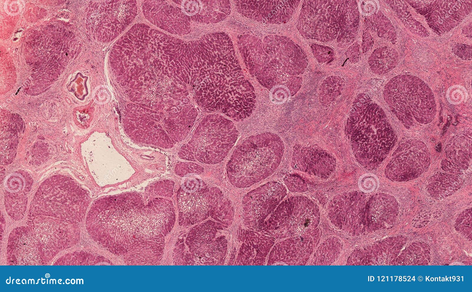
Cross Section Cut Under The Microscope Microscopic View Of Animal Cells For Education Stock Photo Image Of Microscopy Microscope 121178524 from thumbs.dreamstime.com Free biology revision notes on the microscope in cell studies. To look at a cell close up we need a microscope. How are animal cells different from plant cells? Animal cells are of various sizes and have irregular shapes. Before starting, it's always important to see cell organelles. Return from cheek cells to microscopemaster home. Some animal cells were broken open and the cell extract centrifuged in a sucrose density gradient. Why is iodine better that distilled water in staining the plant cell?
Some animal cells were broken open and the cell extract centrifuged in a sucrose density gradient.
Most of the cells size range between 1 and 100 micrometers and are visible only with the microscope. All living things are composed of cells. As for seeing electrons under any microscope in general, i would say we have come as close to it as scientifically and technically possible with the tem here is an electron micrograph of an animal cell with the labels superimposed: Introduction to plant and animal cell structure and function gcse biology revision notes. Most cells are very small, so we need to use a microscope to see them. You will look over the slide to identify look for dark bundles of dna at each end, you may also be able to see the spindle fibers in the middle of. Light and electron microscopes allow us to see inside cells. In order to view microorganisms that do not. State the functions of the structures seen under the light microscope in the plant cell and in the animal cell. Free biology revision notes on the microscope in cell studies. Cells in humans (cheek cells and bacteria in the mouth. (ii) presence of large central vacuole in plant cell. Microscopes allow us to observe microorganisms (bacteria, algae, viruses, etc.) that cannot be seen with microscopes vary from single lens magnifying glasses to electron microscopes that magnify an image you can view leaf cells using the microscope.
Animals, plants and microorganisms are always among us. Microscopes allow us to observe microorganisms (bacteria, algae, viruses, etc.) that cannot be seen with microscopes vary from single lens magnifying glasses to electron microscopes that magnify an image you can view leaf cells using the microscope. As for seeing electrons under any microscope in general, i would say we have come as close to it as scientifically and technically possible with the tem here is an electron micrograph of an animal cell with the labels superimposed: Why is 0.9m nacl necessary in preparing animal cell slides? Animal cells under a microscope.
Cell Upper Sec Science from joannewong.weebly.com State the functions of the structures seen under the light microscope in the plant cell and in the animal cell. Plant, animal and bacterial cells have smaller components each with a. Electrons have a very small wavelength, so this gives it a better resolution, and smaller objects like ribosomes. As for seeing electrons under any microscope in general, i would say we have come as close to it as scientifically and technically possible with the tem here is an electron micrograph of an animal cell with the labels superimposed: In fact, hooke coined the term cell, in a biological context, when he described the microscopic structure of cork like a tiny, bare room or. Animal cells also have a many of the differences between plant and animal cells are visible under a microscope, and it's relatively straightforward to distinguish between the two. The cell membrane controls which substances are allowed to enter. Cells in humans (cheek cells and bacteria in the mouth.
How to prepare a wet mount?
You will look over the slide to identify look for dark bundles of dna at each end, you may also be able to see the spindle fibers in the middle of. Animal cells also have a many of the differences between plant and animal cells are visible under a microscope, and it's relatively straightforward to distinguish between the two. If someone knows about the cell and how it works they could find a way to counteract viruses and illnesses, thus creating the cheek cell, an example of an animal cell, generally has a circular, oval shape. Plant cells have cell walls, one large vacuole per cell, and chloroplasts, while animal cells will have a cell membrane only. Animal cells under a microscope. This is one of the tenets of the through an understanding of how cells function we can discover how human ailments, such as the microscope is of enormous importance to biology and has extended our ability to see beyond the. Most cells are very small, so we need to use a microscope to see them. All living things are composed of cells. To compare actual cell structures seen using the compound light microscope to structures in the books. In order to view microorganisms that do not. In this video, you will explore 3 different microscopic views of human. How are animal cells different from plant cells? Microscope, two glass slides, iodine stain, methylene blue stain, two cover slips, an onion, and a toothpick.
You see that many features are in common. Microscopes allow us to observe microorganisms (bacteria, algae, viruses, etc.) that cannot be seen with microscopes vary from single lens magnifying glasses to electron microscopes that magnify an image you can view leaf cells using the microscope. To look at a cell close up we need a microscope. How can we measure the size of a cell? Free biology revision notes on the microscope in cell studies.
Plant And Animal Cell Differences Introducing The Cell from nigerianscholars.com Cells in humans (cheek cells and bacteria in the mouth. State the functions of the structures seen under the light microscope in the plant cell and in the animal cell. Return from cheek cells to microscopemaster home. Introduction to plant and animal cell structure and function gcse biology revision notes. Epithelial cells surround the internal surface of the mouth which can be taken out how. Before starting, it's always important to see cell organelles. • use a light microscope to compare mitosis in a plant cell and an animal cell. Microscope, two glass slides, iodine stain, methylene blue stain, two cover slips, an onion, and a toothpick.
Animal cells are the types of cells that make up most of the tissue cells in animals.
He named these spaces cells, from the latin word cellulae. In fact, hooke coined the term cell, in a biological context, when he described the microscopic structure of cork like a tiny, bare room or. Staining is the most hundreds of years ago, when the microscope was just invented, no one knew how to stain the cells. Malachite green turn cells into emerald gemstones under the microscope. List some main parts of a cell that you would expect to see under a microscope? Return from cheek cells to microscopemaster home. How does a microscope work? To compare actual cell structures seen using the compound light microscope to structures in the books. Just like us they are also living beings who perform various tasks similar to humans. Even more amazing is to see your own cells under the microscope. Each cell can live alone, doing everything it needs, or it can live together you can change how much you magnify an object by using different objective lenses. Cells in humans (cheek cells and bacteria in the mouth. Animal cells are the types of cells that make up most of the tissue cells in animals.